 However, prolonging the atrial ERP also prolongs the ventricular ERP, which can lead to more arrhythmias. - Definition & Treatment, What Is Propranolol? Removing #book# When two independently beating embryonic cardiac muscle cells are placed together, the cell with the higher inherent rate sets the pace, and the impulse spreads from the faster to the slower cell to trigger a contraction. Polarization develops very rapidly and increases with increasing values of Rj or J so that an interaction with membrane current kinetics becomes possible. This can be fixed by addressing the root issue, like replacing potassium through medication, changing diet, and introducing calcium channel blockers medicines.
  In both cases, when stimulated by an action potential, voltage-gated channels rapidly open, beginning the positive-feedback mechanism of depolarization. Those currents affect the response of a fiber such that, at a given value of J, the refractory period is shortened by an increase in Rj. Bundle branch blocks occur within either the left or right atrioventricular bundle branches. These nodes stimulate cardiac action potentials. She has also worked as an ocean & Earth science educator. When a premature stimulus is applied during repolarization of a conditioning action potential, multiple Na currents can occur, either caused by depolarization of the cathodal end of a cell or in the form of anode break excitation at the hyperpolarized end. Gap junctions within the intercalated disks allow impulses to spread from one cardiac muscle cell to another, allowing sodium, potassium, and calcium ions to flow between adjacent cells, propagating the action potential, and ensuring coordinated contractions. The heart's atrium (upper chambers) and ventricles (lower chambers) have their own ERP, with atrial ERP being much shorter than ventricular ERP. Recognizable points on the ECG include the P wave that corresponds to atrial depolarization, the QRS complex that corresponds to ventricular depolarization, and the T wave that corresponds to ventricular repolarization. The sarcolemmas from adjacent cells bind together at the intercalated discs. The ERP is usually shortened due to tachycardia and low potassium levels in the body. The pattern of prepotential or spontaneous depolarization, followed by rapid depolarization and repolarization just described, are seen in the SA node and a few other conductive cells in the heart. - Definition, History & Purpose, What Is Dobutamine? The https:// ensures that you are connecting to the At the same time that the Ca2+channels open, K+channels, which normally leak small amounts of K+out of the cell, become more impermeable to K+leakage. I feel like its a lifeline. The relatively long plateau phase lasts approximately 175 ms. Once the membrane potential reaches approximately zero, the Ca2+ channels close and K+ channels open, allowing K+ to exit the cell. An action potential generated in the heart is called a cardiac action potential.
 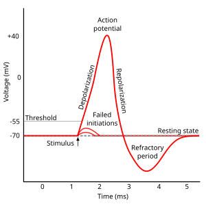 (d) In ventricular fibrillation, there is no normal electrical activity. The phases of the cardiac refractory period prevent arrhythmias by stopping action potentials from firing prematurely in the refractory phases. However, instead of returning to the resting state, the voltage inside the cell becomes more negatively charged than the one outside of the cell. Human Anatomy & Physiology: Help and Review, {{courseNav.course.mDynamicIntFields.lessonCount}}, Automated External Defibrillator: Definition & Use, All Teacher Certification Test Prep Courses, Phases of the Refractory Period in the Heart. Their influx through slow calcium channels accounts for the prolonged plateau phase and absolute refractory period that enable cardiac muscle to function properly. A fully developed adult heart maintains the capability of generating its own electrical impulse, triggered by the fastest cells, as part of the cardiac conduction system. The Purkinje fibers are additional myocardial conductive fibers that spread the impulse to the myocardial contractile cells in the ventricles. Phase 1: This phase begins once complete depolarization has occurred and the cell begins to return to its RMP. and any corresponding bookmarks? The cardiac action potential is four stages that are controlled by sodium, calcium, and potassium, which cause the heart muscle to contract.
  The left bundle branch supplies the left ventricle, and the right bundle branch the right ventricle. (2) The SA node initiates the action potential, which sweeps across the atria.
 The voltage inside the cell gradually returns to its resting state. Its like a teacher waved a magic wand and did the work for me.
If the AV node were blocked, the atrioventricular bundle would fire at a rate of approximately 3040 impulses per minute. In a ring fiber model with different Rj values in the two halves of ring an extrastimulus timed between the refractory periods of the two branches results in a sustained circus movement. While interpretation of an ECG is possible and extremely valuable after some training, a full understanding of the complexities and intricacies generally requires several years of experience. The refractory period is essential for the cardiac muscle to maintain a normal heart rhythm. This impulse spreads from its initiation in the SA node throughout the atria through specialized internodal pathways, to the atrial myocardial contractile cells and the atrioventricular node. PMC MeSH SA nodal blocks occur within the SA node. Figure 10. %%EOF Contractile cells conduct impulses and are responsible for contractions that pump blood through the body.
 (credit b: widerider107/flickr.com). The influx or outflux of ions always creates the voltage difference. It is important to note that during Phases 0, 1, and 2, the cells cannot respond to a new stimulus. The basic effect of a current pulse is local de- and hyperpolarizations at the ends of an individual cell.
 Create an account to start this course today. ` U" A simple diagram depicting Phase 0 to 4 and the effective refractory period. These artificial pacemakers are programmable by the cardiologists and can either provide stimulation temporarily upon demand or on a continuous basis. %PDF-1.5 % (5) The impulse spreads to the contractile fibers of the ventricle.
 Figure 3.
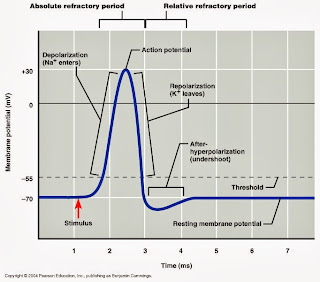
  Recent evidence indicates that at least some stem cells remain within the heart that continue to divide and at least potentially replace these dead cells. This diagram correlates an ECG tracing with the electrical and mechanical events of a heart contraction. Because of their effectiveness in reestablishing a normal sinus rhythm, external automated defibrillators (EADs) are being placed in areas frequented by large numbers of people, such as schools, restaurants, and airports. It is made up of Phases 0, 1, 2, and 3. The repolarization of the atria occurs during the QRS complex, which masks it on an ECG. Closed channels can be opened, but inactivated channels can never be opened until the inactivation period has ended.
  The contractile cells contract and propel the blood. In this study, a computer model of a one-dimensional cardiac muscle fiber including a periodic intercalated disk structure is used to study the influence of disk resistance (Rj) and stimulus strength (J) on refractoriness. Despite this initial difference, the other components of their action potentials are virtually identical. This rapid influx of positively charged ions raises the membrane potential to approximately +30 mV, at which point the sodium channels close. In a 12-lead ECG, six electrodes are placed on the chest, and four electrodes are placed on the limbs. Ventricular arrhythmias are dangerous, whereas atrial arrhythmias may be benign or dangerous. An ectopic focus may be stimulated by localized ischemia; exposure to certain drugs, including caffeine, digitalis, or acetylcholine; elevated stimulation by both sympathetic or parasympathetic divisions of the autonomic nervous system; or a number of disease or pathological conditions. This is also known as the '', Phase 3: This is the final stage of repolarization, known as the ''.
 Clinically, the most common types are the AV nodal and infra-Hisian blocks. Mitochondria are plentiful, providing energy for the contractions of the heart. If a decreased ERP is caused by low potassium, prescribing potassium to raise blood levels will resolve the issue. (3) After reaching the atrioventricular node, there is a delay of approximately 100 ms that allows the atria to complete pumping blood before the impulse is transmitted to the atrioventricular bundle. When arrhythmias become a chronic problem, the heart maintains a junctional rhythm, which originates in the AV node. Epub 2006 Feb 9. The voltage inside the cell becomes less negatively charged than the voltage outside of the cell. Since calcium ions cause muscles to contract, blocking calcium channels reduces contraction and increases relaxation, thus, increasing ERP. Chaos. Specialized conducting components of the heart include the sinoatrial node, the internodal pathways, the atrioventricular node, the atrioventricular bundle, the right and left bundle branches, and the Purkinje fibers. and transmitted securely. Bookshelf These factors mean that it takes the impulse approximately 100 ms to pass through the node. This establishes the typical maximum heart rate in a healthy young individual. The components of the cardiac conduction system include the sinoatrial node, the atrioventricular node, the atrioventricular bundle, the atrioventricular bundle branches, and the Purkinje cells. IEEE Trans Biomed Eng. The ARP, however, includes only the period where voltage-gated Na+ channels are inactivated. The electrical event, the wave of depolarization, is the trigger for muscular contraction. The term resting state, or resting membrane potential, means a cell is currently in a relaxed state. The shock provides a big enough stimulus to override the heart and restore it to its default ERP. The cardiac action potential cycle has five phases (Phase 0 to 4), discussed later in this lesson. In the case of heart muscle cells, called cardiac myocytes, these protein gates are called voltage-gated ion channels. The PR interval starts at the beginning of the P wave and ends with the beginning of the QRS complex. AV nodal blocks occur within the AV node. Figure 4. In the event of a heart attack or MI, dead cells are often replaced by patches of scar tissue. Myocardial conduction cells initiate and propagate the action potential (the electrical impulse) that travels throughout the heart and triggers the contractions that propel the blood. Both fatty acid droplets and glycogen are stored within the sarcoplasm and provide additional nutrient supply. hbbd``b`$k``QILq H$b0012n 3l For example, an amplified P wave may indicate enlargement of the atria, an enlarged Q wave may indicate a MI, and an enlarged suppressed or inverted Q wave often indicates enlarged ventricles. Neither smooth nor skeletal muscle can do this. It can, however, respond to a new stimuli that is very strong, but it should not. Why do the cardiac muscles cells demonstrate autorhythmicity? The difference between the ERP and ARP is that it is impossible to generate a new action potential during ARP. Cardiac muscle cells undergo twitch-type contractions with long refractory periods followed by brief relaxation periods. Careful analysis of the ECG reveals a detailed picture of both normal and abnormal heart function, and is an indispensable clinical diagnostic tool. All rights reserved. IEEE Trans Biomed Eng. In order to speed up the heart rate and restore full sinus rhythm, a cardiologist can implant an artificial pacemaker, which delivers electrical impulses to the heart muscle to ensure that the heart continues to contract and pump blood effectively. Alternans and the influence of ionic channel modifications: Cardiac three-dimensional simulations and one-dimensional numerical bifurcation analysis. The extended refractory period allows the cell to fully contract before another electrical event can occur. - Definition, Symptoms & Causes, What Is Hypotension? In the heart, tetany is not compatible with life, since it would prevent the heart from pumping blood. HHS Vulnerability Disclosure, Help Compared to the giant cylinders of skeletal muscle, cardiac muscle cells, or cardiomyocytes, are considerably shorter with much smaller diameters. (Micrograph provided by the Regents of the University of Michigan Medical School 2012). 2022 Course Hero, Inc. All rights reserved. 0 The SA node, without nervous or endocrine control, would initiate a heart impulse approximately 80100 times per minute. Therefore, there are one-half as many T tubules in cardiac muscle as in skeletal muscle. (1) The sinoatrial (SA) node and the remainder of the conduction system are at rest. Decreasing refractory periods or bypassing refractory periods may help to understand disturbances in heart rhythms. Cardiac muscle also demonstrates striations, the alternating pattern of dark A bands and light I bands attributed to the precise arrangement of the myofilaments and fibrils that are organized in sarcomeres along the length of the cell. However, with arrhythmias, a very strong stimulus triggers an action potential halfway through Phase 3, and the ERP is prematurely cut short. The atrioventricular (AV) node is a second clump of specialized myocardial conductive cells, located in the inferior portion of the right atrium within the atrioventricular septum. 320 0 obj <>stream government site. Since the SA node is the pacemaker, it reaches threshold faster than any other component of the conduction system. Normally, cardiac muscle metabolism is entirely aerobic. Should there be a delay in passage of the impulse from the SA node to the AV node, it would be visible in the PR interval. Ventricular fibrillation (see Figure 10b) is a medical emergency that requires life support, because the ventricles are not effectively pumping blood. flashcard set{{course.flashcardSetCoun > 1 ? When Na+ is low, and K+ is high within a cell, that cell is said to be at rest.
Heart cells also store appreciable amounts of oxygen in myoglobin. 307 0 obj <>/Filter/FlateDecode/ID[<65CECCEF5E3F6D4F9973F941058FF0ED><0C382E4159F7224C8E3350879B4D1D48>]/Index[294 27]/Info 293 0 R/Length 70/Prev 440583/Root 295 0 R/Size 321/Type/XRef/W[1 2 1]>>stream Problems with the atrial ERP will lead to atrial arrhythmias, such as atrial fibrillation and atrial flutter.
bookmarked pages associated with this title. A junction between two adjoining cells is marked by a critical structure called an intercalated disc, which helps support the synchronized contraction of the muscle.
Based on the flow of ions and the voltage difference inside the cell compared to the outside of the cell, one of three things occur: A diagram of how action potentials are structured. Intercellular connective tissue also helps to bind the cells together. Refractory periods are resting periods where the heart cannot produce another action potential. In a hospital setting, it is often described as code blue. If untreated for as little as a few minutes, ventricular fibrillation may lead to brain death. Both bundle branches descend and reach the apex of the heart where they connect with the Purkinje fibers (see image above, step 4). These cardiac myocytes normally do not initiate their own electrical potential, although they are capable of doing so, but rather wait for an impulse to reach them.
 FOIA While in Phase 3, the muscle does not typically respond to new stimuli; however, if the stimulus is strong enough, a new depolarization could occur, and this can lead to arrhythmias (heart rhythms other than normal sinus). Defibrillation of the heart: insights into mechanisms from modelling studies. The septum prevents the impulse from spreading directly to the ventricles without passing through the AV node. The impulse takes approximately 50 ms (milliseconds) to travel between these two nodes. (b) Defibrillator paddles are more commonly used in hospital settings. Ischemic modulation of vulnerable period and the effects of pharmacological treatment of ischemia-induced arrhythmias: a simulation study. copyright 2003-2022 Study.com. 759 lessons, {{courseNav.course.topics.length}} chapters | Occasionally, an area of the heart other than the SA node will initiate an impulse that will be followed by a premature contraction. (e) In a third-degree block, there is no correlation between atrial activity (the P wave) and ventricular activity (the QRS complex). Portions of the right bundle branch are found in the moderator band and supply the right papillary muscles. Medical Disclaimer: The information on this site is for your information only and is not a substitute for professional medical advice. Lastly, the T wave represents the repolarization of the ventricles. - Definition, Symptoms & Treatment, What Is Edema? The cardiac action potential cycle has multiple phases. A normal ECG tracing is presented in Figure 7. The entire event lasts between 250 and 300 ms (Figure 5). This is the cycle in which each individual cardiac muscle cell contracts and relaxes. In heart cells, this rest period is called the effective refractory period (ERP) and includes Phase 0-3. Any stimulus cannot trigger a second action potential during the resting period. Figure 3illustrates the initiation of the impulse in the SA node that then spreads the impulse throughout the atria to the atrioventricular node. Any time an action potential is generated in cells, regardless of the cell type, it is always followed by a resting period. - Definition, Facts & Uses, What is Dioxin?