8798). Sleep Medicine Reviews, 14(4), 219226. In M. S. Gazzaniga (Ed. Consciousness and Cognition, 20(4), 998 1008. doi: 10.1016/j.concog.2010.10.005, Doricchi, F., & Violani, C. (1992). Fisher, C. (1973). Mxico: Planeta.
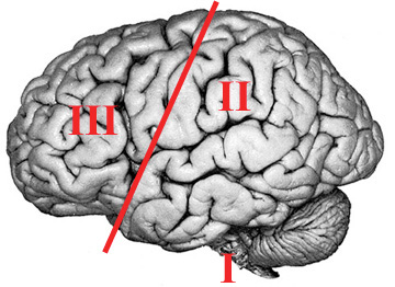
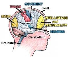
 In general, the prefrontal lobe has been associated with selection functions, programming, and direction of behavioral planning, and impulse inhibition, as well as critical and reflexive thought (Cummings, 1995; Luria, 1974; Tsvetkova, 1996). Electroencephalography and Clinical Neurophysiology, 35(2), 193198. The activation of the supplementary motor area (Brodmanns area 6) and primary motor area (Brodmanns area 4) produces a programming and activation of a sequence of corporal movements during the oneiric content; but said activation remains on a representational level, because an inhibition occurs in the caudal region of locus coeruleus located in the pons of the brain stem (Unit 1) due to hyperpolarization of the motoneurons in the spinal cord. neuropsychological Such research has been examined here. Brain, 120(7), 11731197. It was not until 1900, however, that Sigmund Freud (1966) published his book The Interpretation of Dreams, which included the first scientific approach to the subject from a purely psychological point of view. In: R. Drucker-Coln, M. Shkurovich, & M. B. Sterman (Eds. It is well known that dreams are difficult to remember in wakefulness (Fisher, 1973). luria alexander B) The Second Unit is formed by the parietal, occipital, and temporal lobes, and is responsible for obtaining, processing, integrating, and storing sensory information from the environment. The content of the dream is bizarre by nature, with bizarre defined as featuring incongruities and discontinuities in the time, space, and the characters that appear in it (Corsi-Cabrera et al., 2003).
 doi: 10.1016/B978-0-12-222340-2.50015-X, Vogel, G. W., Vogel, F., McAbee, R. S., & Thurmond, A. J. For example, Pea-Casanova et al. New Jersey: Psychology Press. Tllez, A.
  We propose that the inhibition of prefrontal lobe functioning and the increase in activity of Unit L during REM sleep can have a cognitive and emotional homeostatic function that is important for good psychological performance during wakefulness. Universidad Autnoma de Nuevo Len (UANL), Monterrey, Mexico, Snchez-Juregui, T. de J. During REM sleep in normal people, there is an increase in the activity of Unit L and a decrease in Unit 3; however, we cannot observe the behavioral effects, due to the activation of the cerebral mechanisms that produce the muscle paralysis that comes with this type of sleep, preventing the body from acting out dreams. It also includes vital cognitive functions such as sustained attention, awareness, and insight (Luria, 1974; Cummings, 1995; Stretton, & Thompson, 2012).
  How can it be proven that the hypo-functioning of the prefrontal lobe and the limbic hyper-functioning during dreaming fulfill a homeostatic need for good psychological functioning during wakefulness? Seeking the criminal element. Calvo, J. M.
In addition, the suggested hypothesis of the homeostatic character of REM sleep favors the idea that the brain works in an inverse way during the state of wakefulness to assist better psychological functioning of the individual.
How can it be proven that the hypo-functioning of the prefrontal lobe and the limbic hyper-functioning during dreaming fulfill a homeostatic need for good psychological functioning during wakefulness? Seeking the criminal element. Calvo, J. M.
In addition, the suggested hypothesis of the homeostatic character of REM sleep favors the idea that the brain works in an inverse way during the state of wakefulness to assist better psychological functioning of the individual. Pea-Casanova, J., Roig-Rovira, T., Bermudez, A., & Tolosa-Sarro, E. Furthermore, both conditions show similar neuropsychological functioning: a hypo-functioning of the frontal lobe and an activation of Unit L. These characteristics impede the schizophrenic patient and the dreamer from organizing their thoughts, integrating them with emotions, and turning them into appropriate actions. Madsen, P. L. (1993).
Thus, the main proposal of this model, is that the characteristics of the oneiric content -that is, the lack of planning and control of critical and coherent thought toward what is dreamt, as well as the ease by which emotional and motivational impulses emerge in dreams basically correspond to an increase in the activity of Unit 1, 2 (with the exception of the PTO region), Unit L, and the medial region of the prefrontal lobe that occurs simultaneously with the inhibition of the dorsolateral and orbital regions of Unit 3 (Figure 1). (1989). The contribution of cerebral hemispheric disease to the understanding of dream type and content. On the other hand, the recollection of dreams becomes interesting.
As a consequence of the activation of Unit 1, Unit 2 is stimulated, generating activation in visual, perceptive-imaginative, auditory, linguistic, spatial, and tactile functions. To answer this question, it would be of great help to describe the phenomenology of dreaming.
During this phase, there is also an increase of electroencephalographic (Rechtschaffen & Kales, 1968) and cerebral metabolic activity, which is equal to or greater than that activity during wakefulness (Braun et al., 1997; Madsen, 1993; Maquet et al., 1996; Sakai, Meyer, Karacan, Derman, & Yamamoto, 1980). Tllez, A. In the case of humans, it is interesting to find a clinical sleep disorder that is similar to the oneiric behavior experimentally induced in cats. Neural correlates of insight in dreaming and psychosis. It has been shown that lucid dreams are characterized by being able to freely remember the circumstances of waking life, to think clearly, and to act deliberately upon reflection, all while experiencing a dream world that seems vividly real (LaBerge, 1990). (1997) found low metabolism in the orbitofrontal and dorsolateral regions of the prefrontal lobe during REM sleep, as well as in the inferior parietal association, and simultaneously, an increase in metabolism in the visual and auditory association areas of Unit 2. Science, 281(5380), 11881191. New perspectives for the study of lucid dreaming: From brain stimulation to philosophical theories of self-consciousness. Cognitive and emotional processes during dreaming: a neuroimaging view. Frith, C. D. (2007). This activity produces a perception of hallucinatory images of various sensory modes, as well as a lack of inhibition, a non-selfreflexive thought process, and a lack of planning and direction of such oneiric images.
doi: 10.1016/S0278-2626(03)00037-X, Cummings, J. L. (1995). Furthermore, there is a lack of control over the course of dream scenes, in which there are often violations of the laws of physics.
Developmental Neuropsychology, 4(3), 199230. Maquet, P., Pters, J. M., Aerts, J., Delfiore, G., Degueldre, C., Luxen, A., & Franck, G. (1996). (1998). Hobson and Stickgold (1995) found that during REM sleep, activation of the brainstem starts in the cholinergic system on a pontine level. ajp.2008.08050721, Berger, R. J. doi: 10.1038/383163a0. It has been proven through fMRI that the degree of activation of the frontal lobe and the para-hippocampal region of the limbic system during the presentation of semantic and visual non-verbal information predicts its subsequent recall, showing the important role of these two structures in memory (Brewer, Zhao, Desmond, Glover, & Gabrieli, 1998; Wagner et al., 1998). (2002). El Cerebro en Accin. Schwartz and Maquet (2002) suggested that the bizarre content of dreaming is similar to certain neuropsychological syndromes that produce visual and spatial agnosia. Answering these and other questions will allow continuing progress in this new and interesting field in the neurosciences: the neuropsychology of dreaming. We can conclude that dreams, as well as cognitive activity in wakefulness, come in various forms and contents.
This researcher proposes this theory in light of the observation that the selective deprivation of REM sleep in animals produces increases in aggressive, sexual, and food-seeking behaviors. Seminars in Neurology, 25(1), 117129. 99133). Right hemispheric mediation of dream visualization: A case study. The emotional brain and sleep: An intimate relationship. luria nebraska neuropsychological
Frontal lobe function in temporal lobe epilepsy. During dreaming, only the limbic region is activated, not the prefrontal; this fact produces a partial or total loss of memory of the oneiric content upon waking up in most people (Figure 1). doi: 10.1176/ajp.146.9.1166, Schwartz, S., & Maquet, P. (2002). Some authors have found that individuals with limbic hyper-function, as indexed by increased scores on the Limbic System Checklist, report more threatening dream content than others (Peterson, Henke, & Hayes, 2002). The first approach toward psychobiological scientific research on the subject of dreaming occurred in 1953 when Aserinsky and Kleitman from the University of Chicago published their research, which stated that sleep with rapid eye movement, known as REM sleep, is frequently associated with dream recall. luria brain working Annals of the New York Academy of Sciences, 769(1), 114. doi: 10.1016/j.eplepsyres.2011.10.009. Sleep deprivation makes us more sensitive to emotional and stress-induced stimuli (Vandekerckhove & Cluydts, 2010). Stretton, J., & Thompson, P. J. M. Bertini (Eds. The Psychologist, 13(12), 618-619. In fact, patients with depression show an increase in the metabolism of the dorsolateral region of the prefrontal lobe during REM sleep instead of the decrease which is observed in subjects without depression. Rechtschaffen, A., & Kales, A. doi: 10.1037/10499-008. Science, 134(3482), 840. doi: 10.1126/science.134.3482.840, Braun, A. R., Balkin, T. J., Wesenten, N. J., Carson, R. E., Varga, M., Baldwin, P., Selbie, S., Belenky, G., & Herscovitch, P. (1997). This process allows for an increase in prefrontal lobe functioning and a decrease of limbic activity throughout the day, allowing better impulse control, planning, and self-regulation of behavior. doi: 10.1176/jnp.14.3.283. Foulkes, D. (1982). Rapid eye movement sleep dreaming is characterized by uncoupled EEG activity between frontal and perceptual cortical regions.
El Cerebro Ejecutivo: Los Lbulos Frontales y la Mente Civilizada.
Madrid: Alianza Editorial. However, there are psychological processes that have received little attention in this field; among them is the process of dreaming. Tllez, A., Tllez, H., Tirado, H., Butcher, E., Railey, C., & Mendoza, M. E. This is because of the cognitive and emotional similarities between them, such as the exaggeration of the emotional activity that contributes the deterioration of rationality and to the lack of selective attention and direction of cognitive knowledge which, besides being grotesque, contains a great quantity of confabulations. neuropsychological cognitive approach Solms, M. (2000). It is well-established that lesions or dysfunction in this area in neuropsychological patients result in uninhibited, impulsive, and bizarre behavior. In his new model to explain depression, Beck (2008) affirms that in patients with depression, there is a hyperactivity of the amygdala that causes an excessive reactivity in the presence of negative events, and hypo-activity of the prefrontal lobe that prevents a proper interpretation of events and counteracts the high activity of the amygdala. We can state that the oneiric craziness of every night is a necessary escape valve permitting the person to act sanely during the state of wakefulness.
functional units unit brain luria four shows system neuropsychology dreaming parietal temporal occipital reticular limbic lobes tem lobe sys frontal (2002). El Cerebro Despierto. Heart rhythm control during sleep. This pattern of brain activity explains the recovery of the executive metacognitive abilities and voluntary control that characterizes lucid dreaming (Dresler et al., 2012; Noreika, Windt, Lenggenhager, & Karim, 2010). luria neuropsychology pioneer This phenomenon has been called oneiric behavior (Jouvet et al., 1981). doi: 10.1080/87565648809540405, emaityt, D., Varoneckas, G., & Sokolov, E. (1984).
Nature, 437(7063), 12201222. 233250). (2014). brain neuropsychology children parts diagram human ph february pm JAMA, 257(13), 17861789. Sleep-related injury in 100 adult patients: A polysomnographic and clinical report.
casebook reitan halstead Toward an etho-ethnology of dreaming. International Journal of Dream Research, 3(1), 3645.
Lurias model of the functional units of the brain and the neuropsychology of dreaming, Universidad Autnoma de Nuevo Len (UANL), Monterrey, Mexico, http://psychologyinrussia.com/volumes/pdf/2016_4/psychology_2016_4_7.pdf. These researchers suggested that this temporary dissociation between the executive and perceptual areas is the cause of the characteristic bizarreness of dreams. Making memories: Brain activity that predicts how well visual experience will be remembered. neuropsychology
doi: 10.1016/0024-3205(89)90021-0. Scientific American, 267(5), 126-133. doi: 10.1038/scientificamerican0992-126, Gibbs, W. W. This patient lost the ability to dream and also showed optic aphasia, optic apraxia, aphasia without agraphia, and color agnosia. Tonus of extrinsic laryngeal muscles during sleep and dreaming.
All of this is due to a lack of critical thinking that can evaluate the coherence, or the lack thereof, of what is happening, so there is a passive and uncritical acceptance of everything that happens (Corsi-Cabrera et al., 2003). doi: 10.1001/jama.1987.
(2) the orbitofrontal region, that relates to the regulation of limbic impulses, as well as (3) the parietal-temporal-occipital (PTO), that is involved in visuo-spatial recognition, symbol processing, and face and object recognition. This leads us to think of the oneiric process as basically a process of confabulation, suggesting that dreaming is a type of normal confabulation that happens every night in a cyclical way, but does not differ much from the confabulatory thoughts of patients with frontal lobe damage. They also suggest that these cognitive strategies during adult dreaming are equivalent to the processes of fantasy, and are far from the reality thought of a young child during wakefulness (Piagets preoperational stage). Sleep Medicine Reviews, 20, 92-99. doi: 10.1016/j.smrv.2014.06.004, Durmer, J. S., & Dinges, D. F. (2005). Lurias Model of the Brains Functional Units can be used to explain the generation of dreams and their characteristics. Science, 118, 273274.
Neurological approaches to the dream problem. (1981) cats and is the result of the activation of Units 1, 2, and especially L, along with the simultaneous inhibition of the prefrontal lobe. neuropsychological luria This can be interpreted as the result of a broad activation of Unit L without a cortical regulation (Buchsbaum et al., 1989). (1995). (1987). Normal human sleep: Regional cerebral hemodynamics. Biologa de los sueos y psicoanlisis. Variations of heart rate during sleep as a function of the sleep cycle.
In I. Karacan (Ed.). That is the reason why Hobson and Stickgold (1995) stated that dreaming represents a model for explaining schizophrenia. The British Journal of Psychiatry, 143(3), 221-231. doi: 10.1192/bjp.142.3.221, LaBerge, S. (1990). dreaming, brain, neuropsychology, functional units, Lurias model. Freudian dream theory today. The pre-frontal lobe is divided into three regions: 1) The dorsomedial region, which is associated with executing functions such as the formulation of goals, working memory, planning, execution of plans, and the self-regulation of behavior; 2) the orbital frontal region, which is related to the inhibition and control of impulses and social tact; and 3) the medial region, which has been related to motivation and the process of thinking what another person is thinking, also known as mentalization, a second order process of representation relevant to social skills (Frith, 2007).
Psychology in Russia. This generates a general muscle paralysis (with the exception of ocular movement), that prevents the dream from becoming an action (Berger, 1961; Jouvet, Sastre, & Sakai, 1981). State of the Art, 2008 - 2022. This disorder is named REM sleep behavior disorder (RBD) and is characterized by the absence of the muscle paralysis which is customary during this stage of sleep, as a result of neurological related disorders.
B., Zhao, Z., Desmond, J. E., Glover, G. H., & Gabrieli, J. D. E.
According to Solms (2000), a renowned researcher in the neuropsychology of dreaming, these data support the essential idea proposed by Freud (1966), who maintained that one of the functions of dreaming was to allow instinctive impulses to emerge (limbic) without the censorship mechanism (dorsolateral and orbital prefrontal regions), thus allowing the attainment of repressed desires in a safe way. In contrast to sleepwalking, which occurs during the slow-wave stage of non-REM sleep and in people who generally behave in a peaceful way, patients with RBD frequently have accidents and carry out physical and verbal assaults on other people during these episodes (Tellez, 1998; Schenck, Bundlie, Patterson, & Mahowald, 1987; Schenck, Milner, Hurwitz, Bundlie, & Mahowald, 1989). Studies with positron emission computerized tomography (PET) have confirmed an increase in the brainstems metabolism (Braun et al., 1997), which generates electroencephalographic and metabolic activation, as well as stimulates of the posterior cortical and subcortical areas, especially the limbic-emotional system.